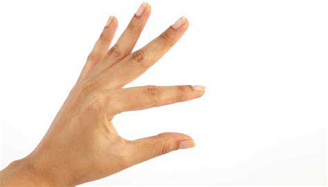
The Science Behind Hand Eye Coordination
Light, the fundamental stimulus for vision, enters the eye through the cornea, which acts as a primary focusing lens. This initial refraction of light is crucial for directing the light rays towards the retina, the light-sensitive tissue lining the back of the eye. The cornea's transparency and curvature are essential characteristics that allow light to pass through unobstructed and begin its journey towards the brain.
The clarity and integrity of the cornea are paramount to visual acuity. Any irregularities or damage to the cornea can significantly impair the quality of the light focused on the retina, leading to blurred or distorted vision. Maintaining healthy corneal function is vital for optimal visual processing.
Photoreceptor Activation: Converting Light to Signals
Upon reaching the retina, light encounters photoreceptor cells, specifically rods and cones. These specialized cells contain photopigments that absorb light energy. This absorption triggers a cascade of biochemical reactions, ultimately converting the light stimulus into electrical signals.
Rods, highly sensitive to low light conditions, are essential for night vision and peripheral perception. Cones, on the other hand, are responsible for color vision and sharp, detailed vision in bright light. The interplay between rods and cones allows for a wide range of visual capabilities.
Signal Transmission Through the Retina
The electrical signals generated by photoreceptors are transmitted through a complex network of retinal neurons. These neurons, including bipolar cells and ganglion cells, process and relay the signals further along the visual pathway. This intricate neural interplay allows for the refinement and organization of visual information.
The Optic Nerve: Relaying the Message to the Brain
The axons of ganglion cells converge to form the optic nerve. This nerve acts as a crucial conduit, carrying the electrical signals from the retina to the brain. The optic nerve is a complex bundle of fibers, responsible for transmitting a vast amount of visual information.
The optic nerve's integrity is vital for transmitting sharp and clear visual information. Damage or interruption to the optic nerve can result in significant visual impairment or even blindness. Protecting the optic nerve is crucial for maintaining visual function.
Thalamic Relay: Processing and Routing
Before reaching the visual cortex, the signals from the optic nerve are relayed through the lateral geniculate nucleus (LGN) of the thalamus. The LGN acts as a crucial processing and relay station, further refining and organizing the visual information. This step ensures the visual signals are appropriately directed to the visual cortex for interpretation.
The thalamus plays a fundamental role in filtering and prioritization of visual information. Its function ensures that the most relevant and crucial visual data reaches the cortex for further processing.
Visual Cortex: Interpreting the World
The visual cortex, located in the occipital lobe of the brain, is the final destination for the visual signals. Here, the complex process of visual perception takes place. The visual cortex interprets the signals from the retina, allowing us to recognize objects, shapes, colors, and movements in our environment. This sophisticated processing allows us to understand the world around us.
The visual cortex is a marvel of biological engineering, enabling us to interpret the vast amount of visual information that constantly bombards our eyes. This intricate process is essential for our interaction with and understanding of the world.
The Feedback Loop: Refining Precision
Understanding the Role of Sensory Input
The feedback loop in hand-eye coordination hinges critically on the constant stream of sensory information. Our eyes provide visual data about the target, its location, and its movement, while proprioceptors in our muscles and joints inform us about the position and movement of our hands. This constant interplay between visual and kinesthetic feedback is vital for refining our actions. Imagine trying to catch a ball – your eyes track its trajectory, sending signals to your brain. Simultaneously, your body registers the position of your hand and arm, allowing you to adjust your movements in real-time. This continuous stream of sensory data is processed and interpreted to create the necessary adjustments, leading to more precise and accurate movements.
The brain doesn't simply react to sensory input; it actively interprets and predicts. This predictive element is crucial for anticipatory movements. For example, when catching a fast-moving ball, the brain anticipates the ball's trajectory and position, initiating the hand and arm movements before the ball reaches the point of contact. This anticipatory aspect of the feedback loop ensures that the hand and eye are in the correct position to achieve the desired outcome, demonstrating a sophisticated interplay between perception and action.
The Refinement Process: Practice and Adaptation
The feedback loop isn't static; it dynamically adapts and refines with practice. Every time we perform a hand-eye coordination task, whether it's hitting a baseball, playing a musical instrument, or simply picking up a cup, we receive feedback about the accuracy and effectiveness of our actions. This feedback allows us to identify areas for improvement and adjust our motor commands to achieve greater precision in subsequent attempts. The more we practice, the more refined the feedback loop becomes, leading to smoother, more accurate movements.
This process of adaptation is also influenced by the complexity of the task. Simple tasks, like catching a stationary ball, may require less complex feedback loops. However, more complex tasks, like playing a sport with multiple moving objects, demand a more sophisticated and adaptive feedback loop. The brain constantly adjusts to the changing demands of the task, refining its predictions and motor commands to optimize performance.
Furthermore, the feedback loop can be influenced by external factors such as fatigue, distractions, and even the environment. These factors can affect the accuracy and speed of the sensory input and the brain's processing time, impacting the overall precision of the hand-eye coordination. Understanding these factors is crucial for optimizing performance in various situations.
Factors Influencing Hand-Eye Coordination
Biological Factors
Several Biological factors play a crucial role in shaping hand-eye coordination. Genetic predisposition undoubtedly contributes to the underlying neural pathways and muscle control necessary for precise movements. Individuals with a family history of high dexterity or athleticism often exhibit superior hand-eye coordination. This suggests a strong genetic component influencing the development of these skills early in life. Furthermore, the structure and development of the brain regions responsible for processing visual information and motor control are intricately linked to the refinement of coordination.
Physiological factors, such as the efficiency of nerve impulses transmitting signals between the brain and the muscles, also contribute significantly. Differences in the speed and accuracy of these signals can affect the precision and responsiveness of hand-eye coordination. Variations in muscle strength and flexibility also influence the ability to perform complex movements with speed and control. In essence, the interplay of genetic predispositions, neural pathways, and physiological characteristics establishes a foundation for the development of hand-eye coordination.
Environmental Influences
The environment significantly impacts the development and refinement of hand-eye coordination. Early childhood experiences, including exposure to stimulating activities and opportunities for practice, are pivotal in fostering these skills. Games and activities that require visual tracking, aiming, or catching, such as throwing a ball or playing catch, provide valuable opportunities for practice and improvement. Consistent engagement in such activities reinforces neural pathways and strengthens the connection between the visual and motor systems.
Cultural and societal factors also play a role. Cultures that emphasize certain skills, such as sports or crafts, might foster more developed hand-eye coordination in their members. Access to appropriate tools and resources, and the encouragement to explore and experiment with these tools, also contribute to the development and enhancement of these abilities.
Practice and Training
The adage practice makes perfect holds true for hand-eye coordination. Regular engagement in activities that require precise hand movements and visual tracking is crucial for improving these skills. The more you practice, the more refined the neural pathways become, leading to enhanced speed, accuracy, and control in coordinating hand and eye movements. Consistent repetition and focused training programs can be highly effective in developing and refining these skills over time.
Age and Development
Hand-eye coordination typically develops in stages throughout childhood. Infants begin with basic reflexes and gradually progress to more complex movements as their nervous system matures. The development of hand-eye coordination is closely tied to overall motor skill development and cognitive maturation. As children grow older, their hand-eye coordination skills typically reach a peak, but ongoing practice and engagement in activities that challenge these skills can maintain and even enhance them throughout life.
Cognitive Factors
Cognitive processes like attention, reaction time, and spatial awareness all significantly influence hand-eye coordination. The ability to focus on visual stimuli, process information quickly, and understand spatial relationships between objects and the body are vital components of effective coordination. Individuals with superior cognitive skills often display better hand-eye coordination, as they can more effectively integrate visual information with motor commands.
Furthermore, the ability to anticipate movements and react accordingly is a critical aspect of hand-eye coordination. This anticipatory skill is often honed through practice and experience, enabling individuals to adjust their hand movements in response to changing visual cues. This adaptability and quick reaction time are crucial for activities ranging from sports to everyday tasks.
Read more about The Science Behind Hand Eye Coordination