Exploring the Structure of Arm Bones
The Radius: A Key Player in Forearm Rotation
The radius, positioned on the thumb side of the forearm, plays a crucial role in the complex movements of the hand and wrist. This long bone extends from the elbow to the wrist and serves as the primary driver for pronation and supination - those essential motions that let us flip our palms upward or downward. What makes the radius particularly fascinating is its unique anatomical structure, featuring a specialized head that interacts smoothly with both the ulna and wrist bones. This design enables everything from firm grips to delicate writing motions.
The Ulna: Providing Stability and Leverage
Running parallel to the radius on the pinky finger side, the ulna acts as the forearm's structural anchor. While slightly longer and more robust than its counterpart, this bone's true value lies in its stabilizing function. The ulna's most distinctive feature is undoubtedly the olecranon process - that prominent bony projection you can feel when you touch your elbow. This anatomical landmark plays a critical role in both the mechanics and limitations of elbow movement.
Articulation and Joint Function: A Seamless Connection
These forearm bones don't work in isolation - they form an intricate partnership with each other and surrounding structures. At the elbow, they connect with the humerus (upper arm bone), while at the wrist they meet the carpal bones. This sophisticated network of joints creates a biological transmission system that converts upper arm power into precise hand movements. Whether you're lifting groceries or performing delicate surgical procedures, this coordinated system makes it possible.
Bone Structure and Composition: A Look at Internal Features
Examining these bones at a microscopic level reveals an engineering marvel. The outer shell consists of dense compact bone that can withstand significant forces, while the interior contains a lattice-like spongy bone that provides shock absorption. This dual-layer design achieves an optimal balance - maximum strength with minimum weight. The exact ratio of these components varies along the bone's length, adapting to different mechanical demands at each section.
Growth and Development: Shaping the Forearm Over Time
During childhood and adolescence, specialized growth plates at each end of these bones orchestrate their lengthening process. These cartilaginous regions gradually transform into solid bone at different rates - typically completing between ages 14-18 in girls and 16-20 in boys. Nutritional factors like calcium and vitamin D intake during these years can significantly influence the bones' final strength and density, with lifelong implications for skeletal health.
Key Articulations: Connecting the Bones
Key Articulations: Defining the Joints
Understanding the articulations, or joints, where arm bones connect is crucial for comprehending the arm's complex movements. The human arm contains several specialized joint types - from the hinge-like elbow to the ball-and-socket shoulder - each engineered for specific movement patterns. This mechanical diversity enables everything from the powerful swing of a hammer to the delicate rotation of a screwdriver.
Humeroulnar Joint: The Hinge of the Forearm
This primary elbow joint functions like a well-oiled door hinge, but with precise biological engineering. The trochlear notch of the ulna fits perfectly into the corresponding groove of the humerus, creating a movement arc of about 140-150 degrees. Ligaments surrounding the joint act like biological seatbelts, preventing dislocation while allowing smooth motion. This design explains why we can lift heavy objects without our elbows bending backward.
Humeroradial Joint: Rotation and Pronation
While the humeroulnar joint handles bending, the humeroradial joint specializes in rotation. The rounded capitulum of the humerus articulates with the concave radial head, creating a joint that's both stable and mobile. This unique combination allows the radius to pivot around the ulna, enabling those essential twisting motions we use countless times daily - from turning doorknobs to stirring coffee.
Radiocarpal Joint: Wrist Articulation
At the wrist, the radius forms the primary connection with the carpal bones in what's essentially a modified ball-and-socket joint. The scaphoid and lunate bones fit into a shallow depression on the radius's distal end, creating remarkable mobility. This joint's design explains why our wrists can move in so many directions while still maintaining enough stability for weight-bearing activities like push-ups.
Proximal Radioulnar Joint: Critical for Forearm Rotation
Located near the elbow, this pivot joint allows the radius to rotate around the stationary ulna. The radial head spins within the radial notch of the ulna, stabilized by the annular ligament. This joint's precision engineering enables the 180-degree rotation that separates human dexterity from most other mammals.
Distal Radioulnar Joint: Completing the Forearm Articulation
At the wrist end, this joint works in concert with its proximal counterpart to complete the forearm's rotational mechanism. The ulnar notch of the radius articulates with the ulnar head, creating a stable platform that still allows rotational movement. This distal joint bears significant load during weight-bearing activities, explaining its robust ligamentous support.
Bone Growth and Development: A Dynamic Process
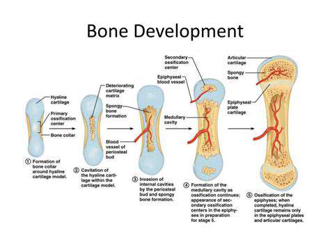
Bone Formation and Ossification
Bone formation, a complex process known as ossification, represents one of nature's most remarkable metamorphoses. In the developing fetus, cartilage models gradually transform into living bone through two distinct pathways. Flat bones like the skull form through intramembranous ossification, where connective tissue transforms directly into bone. Long bones like the radius and ulna develop through endochondral ossification, where cartilage serves as a temporary scaffold that's progressively replaced by bone tissue.
Factors Influencing Bone Growth
Multiple biological systems interact to regulate skeletal development. While genetics provide the blueprint, environmental factors significantly influence the final construction. Adequate nutrition - particularly protein, calcium, phosphorus, and vitamin D - serves as the building materials. Hormones act as the construction foremen, with growth hormone driving overall growth and sex hormones determining the timing of growth plate closure.
Growth Plate Activity and Closure
The growth plates (epiphyseal plates) represent the engine rooms of bone lengthening. These specialized cartilaginous regions contain stacked columns of chondrocytes that multiply, mature, and eventually become replaced by bone. The timing of growth plate closure varies significantly by bone, sex, and individual genetic factors, ultimately determining adult height and limb proportions.
Bone Remodeling and Maintenance
Even after growth ceases, bones remain dynamic tissues through constant remodeling. Specialized cells called osteoclasts tunnel through old bone, while osteoblasts follow behind laying down new matrix. This continuous renewal process allows bones to adapt to mechanical stresses, repair microdamage, and participate in mineral homeostasis throughout life.
Clinical Significance: Conditions Affecting Arm Bones
Clinical Significance of Cardiovascular Conditions
These conditions are a leading cause of morbidity and mortality worldwide, yet many remain preventable through lifestyle modifications. The economic burden extends beyond healthcare costs to include lost productivity and reduced quality of life. Public health initiatives focusing on risk factor reduction have demonstrated significant success in lowering cardiovascular disease rates in many populations.
Hypertension: A Silent Killer
Often called the silent killer because it frequently presents no symptoms, high blood pressure damages blood vessels throughout the body. The continuous excessive pressure causes microscopic tears in arterial walls that become sites for plaque accumulation. Modern treatment approaches emphasize a combination of lifestyle changes and carefully selected medications tailored to individual patient profiles.
Coronary Artery Disease (CAD): A Narrowing of the Arteries
The development of CAD represents a decades-long process beginning as early as adolescence. Plaque buildup progresses through distinct stages, from fatty streaks to complex lesions that can rupture. Contemporary treatment strategies focus not just on opening blocked arteries, but on comprehensive plaque stabilization and regression through aggressive risk factor management.
Heart Failure: A Progressive Condition
Modern classification systems recognize heart failure as a spectrum rather than a single condition, with preserved or reduced ejection fraction subtypes requiring different treatment approaches. Recent therapeutic advances have transformed what was once considered a uniformly terminal condition into a manageable chronic disease for many patients.
Stroke: A Brain-Damaging Event
The concept of time is brain underscores the urgency of stroke treatment, with each minute of delay resulting in the loss of millions of neurons. Advanced imaging techniques now allow clinicians to identify salvageable brain tissue beyond the traditional treatment window in selected patients.
Peripheral Artery Disease (PAD): Limb-Threatening Condition
PAD serves as a marker for systemic atherosclerosis, with many patients having concurrent coronary or cerebrovascular disease. Modern endovascular techniques have revolutionized treatment, offering minimally invasive options for limb salvage in many cases where amputation was previously inevitable.
Atherosclerosis: The Underlying Cause
Current research focuses on understanding the inflammatory components of atherosclerosis and developing targeted therapies. Emerging evidence suggests that plaque composition may be more important than degree of stenosis in predicting clinical events.
Read more about Exploring the Structure of Arm Bones


Hot Recommendations
- The Impact of the Digital Age on Hand Function
- The Role of Hands in Agricultural Innovation
- The Impact of Technology on Hand Artistry
- The Importance of Hand Care for Artists
- How Hand Control Enhances Robotic Surgery
- The Impact of Hand Strength on Physical Labor
- How Handwriting Influences Cognitive Development
- The Impact of Environmental Factors on Hand Health
- The Power of Hands in Building Community
- The Importance of Ergonomics in Hand Health